


Diseases and conditions
Want to know what dermatologists tell their patients about managing conditions that affect the skin, hair, or nails? You’ll find their expertise and insight here.

During the coronavirus pandemic, the AAD's Coronavirus Resource Center will help you find information about changes in dermatology and how you can continue to care for your skin, hair, and nails.

When you have acne, you need two things for clearer skin. The first is acne-friendly skin care. You also need the right treatment. You’ll find the skin care and treatment advice that dermatologists give their patients here.

Here you’ll find information about different types of eczema. This dermatologist-reviewed information will help you better understand and manage the type of eczema you have. You’ll also learn when to see a board-certified dermatologist.

Hair loss can be difficult to talk about. That's why the AAD created the Hair Loss Resource Center. Here, you'll find dermatologists’ insight that can help you discover what steps to take. In many cases, hair loss can be stopped or treated.

Psoriasis is often a lifelong condition. Here, you’ll find dermatologists' insight that can help you diminish psoriasis, reduce flare-ups, and feel better.

Rosacea is a common skin condition that dermatologists frequently treat. Here, you’ll find dermatologists’ insight that can help you with the redness, acne-like breakouts, sensitive skin, along with other signs and symptoms.

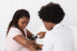
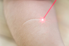
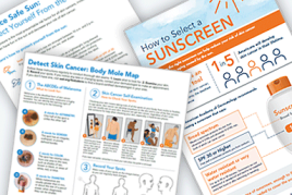
Skin cancer is the most common cancer in the United States. It’s also one of the most preventable and highly treatable when found early. Here, you'll find dermatologists' expertise to prevent skin cancer, along with information for treatment.

Find information about diseases that affect the skin, hair, or nails in people of all skin tones.

A video gallery of what dermatologists tell their patients about managing diseases and conditions that affect the skin, hair, and nails.